Nanostructure promotes growth of new blood vessels, mimics natural protein
Tissue deprived of oxygen (ischemia) is a serious health condition that can lead to damaged heart tissue following a heart attack and, in the case of peripheral arterial disease in limbs, amputation, particularly in diabetic patients.
Northwestern University researchers have developed a novel nanostructure that promotes the growth of new blood vessels and shows promise as a therapy for conditions where increased blood flow is needed to supply oxygen to tissue.
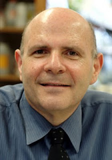
"An important goal in regenerative medicine is the ability to grow blood vessels on demand," said Samuel I. Stupp, Board of Trustees Professor of Chemistry, Materials Science and Engineering, and Medicine. "Enhancing blood flow at a given site is important where blood vessels are constricted or obstructed as well as in organ transplantation where blood is needed to feed the cells properly."
Stupp led the study that will be published the week of Aug. 1 by the Proceedings of the National Academy of Sciences (PNAS).
Stupp and his team designed an artificial structure that, like the natural protein it mimics, can trigger a cascade of complex events that promote the growth of new blood vessels. The protein the nanostructure mimics is called vascular endothelial growth factor, or VEGF.
The nanostructure, however, exhibits important advantages over VEGF: it remains in the tissue where it is needed for a longer period of time; it is easily injected as a liquid to the tissue; and, relative to the protein, it is inexpensive to produce. (VEGF was tested in human clinical trials but without good results, possibly due to it remaining in the tissue for only a few hours.)
"We approached this as an engineering problem," said first author Matthew Webber, a doctoral student in Stupp's research group at the Institute for BioNanotechnology in Medicine (IBNAM). "To be able to design and create a small molecule that can assemble into nanostructures that function therapeutically is rewarding."
Stupp and his team created a nanostructure in the form of a fiber that displays on its surface a high density of peptides (potentially hundreds of thousands) per fiber. The peptides mimic the biological effect of VEGF, initiating the signaling process in cells that leads to blood vessel growth.
The extremely large number of active peptides results in a very potent therapeutic, and the size and stability of the nanofiber ensure the structure is retained longer in the tissue after injection.
After developing the nanostructure, Stupp and Webber teamed up with Losordo to test the nanostructures in vivo.
The researchers used an animal model of peripheral arterial disease and demonstrated the effectiveness of the nanofiber in treating the condition. In animals whose limbs were restricted to only 5 to 10 percent of normal blood flow, treatment with the nanofiber resulted in blood flow being restored to 75 to 80 percent of normal levels.
Treatment with the peptide alone did not produce the same therapeutic effect; the nanostructure was needed to display the peptides to produce results.
"Using simple chemistry, we have produced an artificial structure by design that can trigger complex events," said Stupp, who is director of IBNAM. "Our nanostructure shows the promise of a general approach to mimicking proteins for broader use in medicine and biotechnology."
The researchers next plan to investigate the protein mimic in a heart attack animal model.
###
The National Institutes of Health supported the research.
The paper is titled "Supramolecular Nanostructures that Mimic VEGF as a Strategy for Ischemic Tissue Repair." In addition to Stupp, Losordo and Webber, other authors of the paper are Jörn Tongers, Christina Newcomb and Katja-Theres Marquardt, of Northwestern University; and Johan Bauersachs, of Hannover Medical School, Hannover, Germany.
Contact: Megan Fellman is the science and engineering editor. Contact her at [email protected] 847-491-3115 Northwestern University
SOURCE
